Take a good look inside…
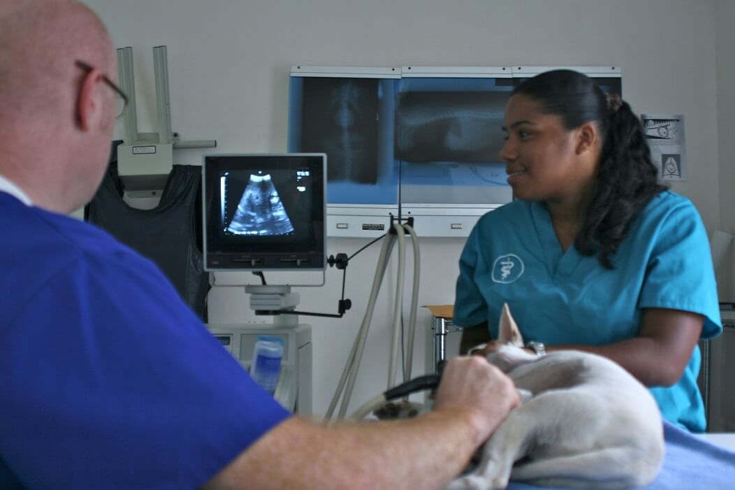
It is amazing what can be found – without surgery – inside the body of a pet, if the right imaging equipment is used and the right person is looking. Our veterinarians are using modern imaging techniques and interpretation procedures to obtain an accurate diagnosis or prescribe a patient-specific treatment plan or simply monitor medical progress.
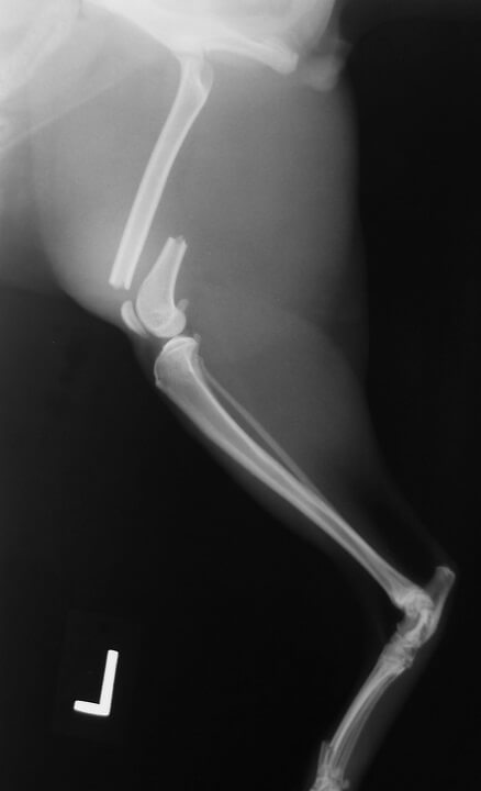
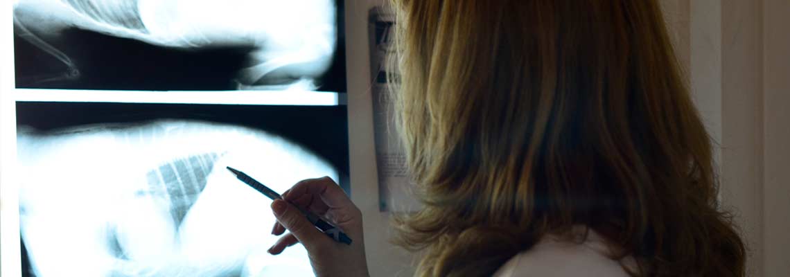
Radiography (xrays) specifically images bone structures, but it can also provide information about many soft tissue densities. As only a 2D image is produced, inevitably your pet will require a minimum of two radiographs to be taken to provide an accurate image of the body part in question and obtain a thorough diagnosis. When radiographs are taken, radiation is produced and therefore there is some risk to personnel. Since some radiographs require manipulating painful or uncomfortable body parts, your pet may receive a small sedative or anesthetic to minimize the time that your pet and the veterinary staff are exposed to the xrays. Again all precautions are implemented to maintain your pet’s safety and comfort.
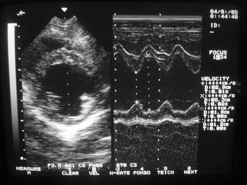
Ultrasound allows many body organs and systems to be visualized, including the liver, gall bladder, spleen, kidney, pancreas and bladder. Ultrasound uses sound waves to image these organs accurately. As this is a painless procedure, we can often perform this technique without the use of sedatives or anesthetics thereby placing minimal burden on your pet’s bodily systems. This is particularly useful for heart conditions or evaluating pets with challenged fitness due to debilitation and disease.